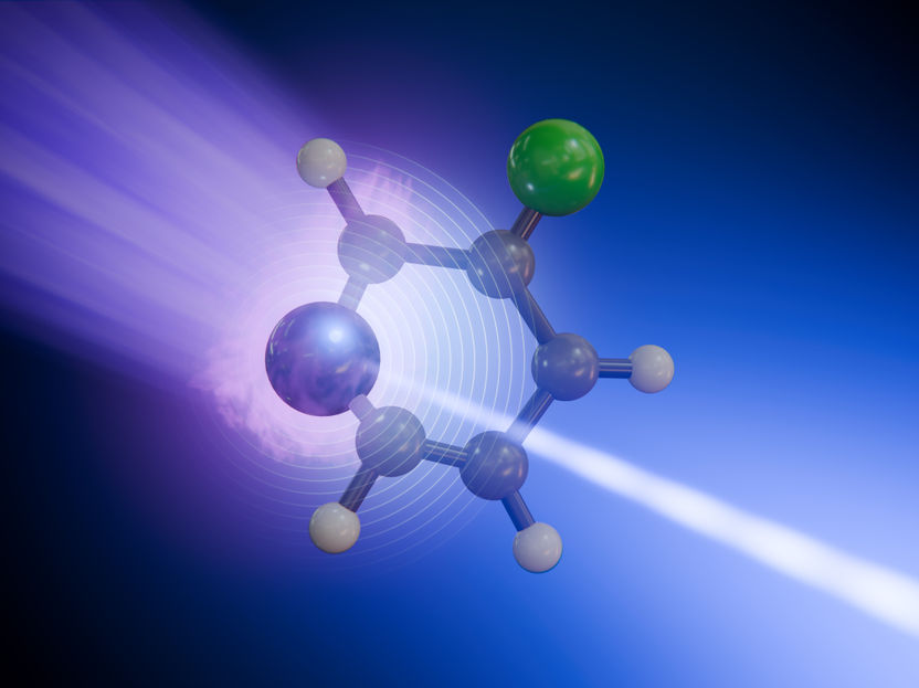
Researchers have captured how a molecule redistributes energy after absorbing light, differentiating the roles of individual atoms in the process.
They used X-ray flashes from the European XFEL to show that different atoms of the same molecule can reveal entirely different aspects of the process.
The study provides clear evidence that excitation by light can enhance an atom’s sensitivity to motion of nearby atoms.
It then passes through a so-called conical intersection: a short-lived but crucial crossing point where movements of electrons and the atoms’ cores become strongly coupled.
An ultraviolet laser pulse first excited the molecules, and a precisely delayed soft X-ray pulse then ionized them by removing deeply bound electrons from either the nitrogen or fluorine atoms.
Researchers have captured how a molecule redistributes energy after absorbing light, differentiating the roles of individual atoms in the process. They used X-ray flashes from the European XFEL to show that different atoms of the same molecule can reveal entirely different aspects of the process. The study provides clear evidence that excitation by light can enhance an atom’s sensitivity to motion of nearby atoms. The new method for following ultrafast chemical reactions at the atomic scale and in real-time can help understanding photostability in DNA, energy flow in light-harvesting materials, and other fundamental processes driven by light.
The team investigated 3-fluoropyridine, a small ring-shaped molecule. When the molecule absorbs light like a short pulse from an ultraviolet laser, it is promoted into an electronically excited state and rapidly distorts out of its original planar shape. It then passes through a so-called conical intersection: a short-lived but crucial crossing point where movements of electrons and the atoms’ cores become strongly coupled. After this point, the molecule returns to the ground state. At that moment, electronic energy is converted into vibrations. The researchers found that this conversion leaves distinct fingerprints at different atomic sites: the fluorine atom acts as a clean marker of vibrational relaxation, while the nitrogen atom, which is more directly involved in the excitation, reflects an intertwined response of electron redistribution and structural motion. “We can now see that not every atomic site tells the same story in the signals we capture from our X-ray pulses,” says Antonio Picón from the Instituto de Ciencia de Materiales de Madrid Consejo Superior de Investigaciones Científicas (ICMM-CSIC), co-author of the study. “Some atoms report where the charge is going, while others reveal how the whole molecule vibrates.”
To observe this process, the team used time-resolved X-ray photoelectron spectroscopy (tr-XPS) at the Small Quantum Systems instrument (SQS) of European XFEL. An ultraviolet laser pulse first excited the molecules, and a precisely delayed soft X-ray pulse then ionized them by removing deeply bound electrons from either the nitrogen or fluorine atoms. By measuring the energy of these emitted electrons at many different time delays, the scientists reconstructed how the local chemical environment evolved over the course of just a couple of picoseconds (trillionths of seconds). To interpret the data, the team developed advanced simulations and models.