The study found that fibroblasts within the tumor microenvironment play a central role in shaping blood vessel formation, oxygen availability, and treatment response.
Anti-angiogenic treatments are of interest since blood vessel growth has been shown to contribute to immune suppression and treatment resistance.
But while anti-angiogenic drugs have shown benefits in lung adenocarcinoma, their efficacy has been limited or have produced toxicity as a treatment for squamous cell carcinoma.
Adenocarcinoma tumors had greater angiogenesis, larger vessel lumens, higher oxygen availability, and less necrosis and hypoxia, while squamous cell carcinomas exhibited poorer blood vessel formation, increased hypoxia, and more necrotic tissue.
In squamous cell carcinoma, however, fibroblasts behaved differently.
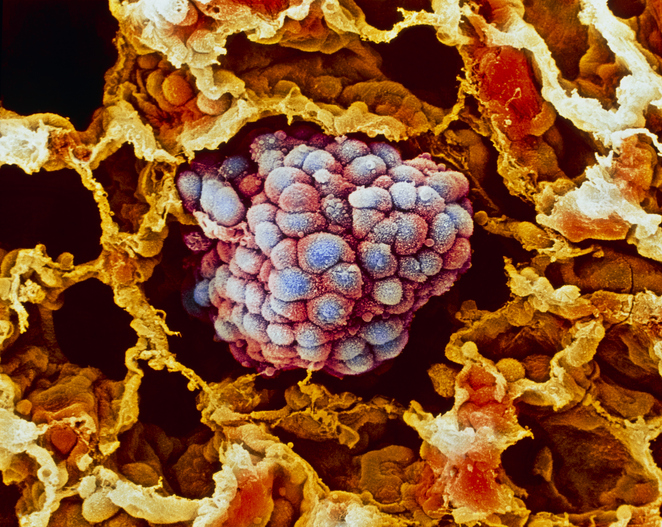
An international collaboration led by researchers at the University of Barcelona, Spain, have identified why the two most common forms of non-small cell lung cancer (NSCLC)—lung adenocarcinoma (LUAD) and squamous cell carcinoma (LUSC)—respond differently to anti-angiogenic therapies that target the blood vessels tumors need to grow. The study found that fibroblasts within the tumor microenvironment play a central role in shaping blood vessel formation, oxygen availability, and treatment response. The findings, published in the journal Cell Death & Disease, could pave the way for treatment decisions that are guided not only by tumor type but also by characteristics of the tumor microenvironment.
“The study reveals that the fibroblast-rich tumor microenvironment is not merely a spectator but a key player that shapes the tumor’s progression. Tumor-associated fibroblasts can influence the vascular network, the availability of oxygen and nutrients and, potentially, also metastatic dissemination and the immune response,” said senior author Jordi Alcaraz, PhD, a professor at the University of Barcelona’s Faculty of Medicine and Health Sciences and researcher at the Institute for Bioengineering of Catalonia.
The new study provides additional evidence that can be used to ascertain the viability of combining anti-angiogenic treatments with immunotherapy. Interest in this potential combination therapy has been growing over the past several years, to address the limited success of immunotherapy alone in treating NSCLC. Anti-angiogenic treatments are of interest since blood vessel growth has been shown to contribute to immune suppression and treatment resistance. But while anti-angiogenic drugs have shown benefits in lung adenocarcinoma, their efficacy has been limited or have produced toxicity as a treatment for squamous cell carcinoma.
“These contrasting outcomes and the limited overall therapeutic benefits attained by anti-angiogenic drugs in LUAD (adenomcarcinoma) underscore the need for a more nuanced understanding of the histotype-dependent complexity of angiogenesis regulation in NSCLC,” the researchers wrote.
For this study, the international team analyzed markers of angiogenesis and hypoxia across multiple patient cohorts. They used a combination of transcriptomic analyses in both lab and animal studies to examined how tumor-associated fibroblasts regulate blood vessel formation in the two major lung cancer subtypes.
The data showed clear differences between the two cancers. Adenocarcinoma tumors had greater angiogenesis, larger vessel lumens, higher oxygen availability, and less necrosis and hypoxia, while squamous cell carcinomas exhibited poorer blood vessel formation, increased hypoxia, and more necrotic tissue.
The investigators determined that fibroblasts from adenocarcinoma tumors produced a pro-angiogenic secretome characterized by elevated levels of vascular endothelial growth factor A (VEGF-A) and tissue inhibitor of metalloproteinases-1 (TIMP-1). “The LUAD-TAF secretome was primed for angiogenesis through SMAD3-dependent overproduction of key regulators, particularly TIMP-1 and VEGF-A,” the researchers wrote adding that “we also uncovered a novel function for TIMP-1 in promoting endothelial cell hyperbranching over basal VEGF signaling.” The findings showed that TIMP-1 amplified VEGF-driven angiogenesis and increased formation of the branching vascular networks commonly associated with tumors.
In squamous cell carcinoma, however, fibroblasts behaved differently. “By contrast,” Alcaraz said, “in squamous cell carcinoma, blood vessel formation is inefficient due to molecular changes in the associated fibroblasts resulting from higher tobacco exposure, resulting in tumors with lower oxygen levels, that is, more hypoxic.”
The findings may also help explain differences in disease progression between the two cancers. Because metastasis depends on tumor cells gaining access to blood vessels, the more developed vascular networks in adenocarcinoma likely play a role to its tendency to spread earlier than squamous cell carcinoma.
These finding could influence new approaches for stratification and treatment selection of patients with NSCLC. Potential biomarkers include the SMAD3/SMAD2 ratio, TIMP-1 levels, VEGF-A expression, and hypoxia-associated fibroblast signatures. The researchers suggest that adenocarcinoma patients may benefit from therapies targeting stromal SMAD3 signaling or TIMP-1, while patients with squamous cell carcinoma may benefit more from treatments designed to counter hypoxia, acidosis, or other features of the tumor microenvironment.
Future research will focus on validating biomarkers such as TIMP-1 in larger patient populations and prospective studies, better understanding the molecular interactions that link SMAD3, VEGF-A, and TIMP-1, and developing treatments that target these pathways. As Alcaraz noted, the challenge now is to identify robust biomarkers, validate them prospectively, and demonstrate that modifying the tumor microenvironment can improve therapeutic responses.