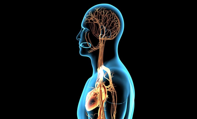
CARDIAC innervation-on-a-chip recreated key neuro-cardiac interactions in a fully human in-vitro model, demonstrating functional communication between neurons and cardiomyocytes.
Axons extended through micro-tunnels linking cortical neurons to sympathetic neurons and sympathetic neurons to cardiomyocytes, creating physical connections that mirrored neurocardiac organisation.
Functional Cardiac Innervation-on-a-Chip ResponsesGene expression and neurotransmitter analyses supported the presence of functional adrenergic and cholinergic signalling pathways within the model.
Subsequent stimulation of sympathetic neurons with acetylcholine further elevated cardiomyocyte beating frequency from 29.19 BPM to 31.42 BPM.
The researchers concluded that this cardiac innervation-on-a-chip platform successfully reproduced physical and functional brain-heart interactions using entirely human cell populations.
CARDIAC innervation-on-a-chip recreated key neuro-cardiac interactions in a fully human in-vitro model, demonstrating functional communication between neurons and cardiomyocytes.
Researchers developed a compartmentalised microfluidic platform that combined human-induced pluripotent stem cell derived cortical neurons, postganglionic sympathetic neurons, and ventricular cardiomyocytes to model the brain-heart axis.
Building A Human Brain-Heart Axis Model
The investigators used a three-compartment microfluidic device known as a 3D3C chip to culture the three cell types in separate but connected environments.
Axons extended through micro-tunnels linking cortical neurons to sympathetic neurons and sympathetic neurons to cardiomyocytes, creating physical connections that mirrored neurocardiac organisation.
Integrated micro-electrode arrays enabled continuous monitoring of electrical activity across the platform.
Over a 21-day culture period, all cell types remained viable and developed characteristic electrophysiological activity.
Cortical neurons and sympathetic neurons displayed increasing spontaneous firing and burst activity, while cardiomyocytes developed stable beating behaviour.
Structural analyses confirmed direct neuronal contacts with cardiomyocytes, including synaptic markers and sympathetic axonal varicosities associated with neurotransmitter release.
Functional Cardiac Innervation-on-a-Chip Responses
Gene expression and neurotransmitter analyses supported the presence of functional adrenergic and cholinergic signalling pathways within the model.
Norepinephrine secretion was detected and was significantly higher than acetylcholine levels in sympathetic neuron and cardiomyocyte compartments, indicating established sympathetic signalling.
To assess functionality, researchers stimulated cortical neurons with kainic acid.
This increased neuronal activity and was followed by a rise in sympathetic neuronal firing and a significant increase in cardiomyocyte beating rate from 27.84 BPM to 29.19 BPM.
Subsequent stimulation of sympathetic neurons with acetylcholine further elevated cardiomyocyte beating frequency from 29.19 BPM to 31.42 BPM.
Increased norepinephrine release was also observed following neuronal stimulation.
Implications For Disease Modelling
Beyond functional connectivity, sympathetic innervation enhanced cardiomyocyte maturation.
Electrophysiological measurements also demonstrated significant changes in field potential duration following neuronal stimulation.
The researchers concluded that this cardiac innervation-on-a-chip platform successfully reproduced physical and functional brain-heart interactions using entirely human cell populations.
By integrating electrophysiological monitoring with targeted neuronal stimulation, the system provides a promising platform for investigating neurocardiac disease mechanisms and evaluating future therapeutic approaches.
Reference
Pesu E et al. Human cardiac innervation-on-a-chip platform recapitulates neurocardiac interactions and supports future disease modeling. Sci Rep. 2026;DOI:10.1038/s41598-026-62182-2
Featured image: PIC4U on Adobe Stock